what structural characteristic gives us the ability to spool the dna
Learning Objectives
- Describe the biochemical structure of deoxyribonucleotides
- Place the base of operations pairs used in the synthesis of deoxyribonucleotides
- Explain why the double helix of DNA is described every bit antiparallel
In Microbial Metabolism, we discussed 3 classes of macromolecules: proteins, lipids, and carbohydrates. In this affiliate, we volition discuss a fourth grade of macromolecules: nucleic acids. Like other macromolecules, nucleic acids are equanimous of monomers, called nucleotides, which are polymerized to course large strands. Each nucleic acrid strand contains certain nucleotides that appear in a certain order inside the strand, called its base of operations sequence. The base of operations sequence of deoxyribonucleic acrid (Deoxyribonucleic acid) is responsible for conveying and retaining the hereditary information in a cell. In Mechanisms of Microbial Genetics, we will discuss in detail the means in which Dna uses its own base sequence to direct its own synthesis, as well every bit the synthesis of RNA and proteins, which, in plow, gives rise to products with various construction and office. In this section, we will discuss the basic construction and office of Dna.
DNA Nucleotides
The building blocks of nucleic acids are nucleotides. Nucleotides that compose Deoxyribonucleic acid are chosen deoxyribonucleotides. The three components of a deoxyribonucleotide are a v-carbon sugar called deoxyribose, a phosphate grouping, and a nitrogenous base, a nitrogen-containing ring structure that is responsible for complementary base pairing between nucleic acid strands (Figure 1). The carbon atoms of the five-carbon deoxyribose are numbered 1ʹ, 2ʹ, 3ʹ, 4ʹ, and 5ʹ (1ʹ is read every bit "one prime"). A nucleoside comprises the five-carbon sugar and nitrogenous base.

Figure 1. (a) Each deoxyribonucleotide is made upwards of a carbohydrate chosen deoxyribose, a phosphate grouping, and a nitrogenous base of operations—in this case, adenine. (b) The v carbons inside deoxyribose are designated equally 1ʹ, 2ʹ, 3ʹ, 4ʹ, and 5ʹ.
The deoxyribonucleotide is named according to the nitrogenous bases (Figure ii). The nitrogenous bases adenine (A) and guanine (Thousand) are the purines; they have a double-band structure with a six-carbon ring fused to a five-carbon ring. The pyrimidines, cytosine (C) and thymine (T), are smaller nitrogenous bases that have just a vi-carbon band structure.

Figure 2. Nitrogenous bases within Deoxyribonucleic acid are categorized into the ii-ringed purines adenine and guanine and the single-ringed pyrimidines cytosine and thymine. Thymine is unique to DNA.
Individual nucleoside triphosphates combine with each other by covalent bonds known as 5ʹ-3ʹ phosphodiester bonds, or linkages whereby the phosphate grouping fastened to the 5ʹ carbon of the sugar of one nucleotide bonds to the hydroxyl group of the 3ʹ carbon of the sugar of the next nucleotide. Phosphodiester bonding between nucleotides forms the sugar-phosphate backbone, the alternate sugar-phosphate structure composing the framework of a nucleic acid strand (Figure 3). During the polymerization process, deoxynucleotide triphosphates (dNTP) are used. To construct the sugar-phosphate backbone, the ii final phosphates are released from the dNTP as a pyrophosphate. The resulting strand of nucleic acid has a complimentary phosphate group at the 5ʹ carbon end and a gratis hydroxyl grouping at the 3ʹ carbon cease. The two unused phosphate groups from the nucleotide triphosphate are released equally pyrophosphate during phosphodiester bond formation. Pyrophosphate is subsequently hydrolyzed, releasing the energy used to drive nucleotide polymerization.
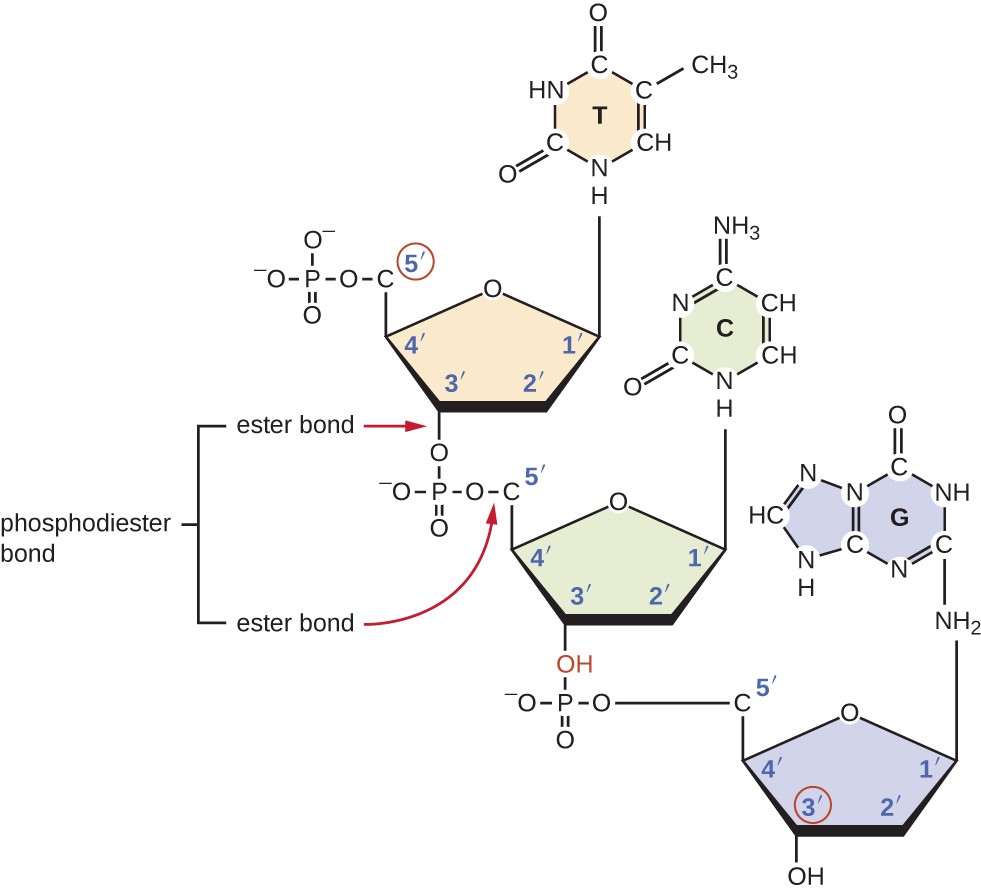
Effigy iii. Phosphodiester bonds form between the phosphate group fastened to the 5ʹ carbon of one nucleotide and the hydroxyl group of the 3ʹ carbon in the next nucleotide, bringing about polymerization of nucleotides in to nucleic acid strands. Annotation the 5ʹ and 3ʹ ends of this nucleic acid strand.
Think well-nigh Information technology
- What is meant past the 5ʹ and 3ʹ ends of a nucleic acrid strand?
Discovering the Double Helix
By the early on 1950s, considerable show had accumulated indicating that Dna was the genetic material of cells, and now the race was on to discover its iii-dimensional structure. Around this time, Austrian biochemist Erwin Chargaff [1] (1905–2002) examined the content of Dna in unlike species and discovered that adenine, thymine, guanine, and cytosine were not found in equal quantities, and that it varied from species to species, simply non between individuals of the same species. He found that the amount of adenine was very close to equaling the amount of thymine, and the amount of cytosine was very shut to equaling the amount of guanine, or A = T and Chiliad = C. These relationships are also known as Chargaff's rules.

Figure 4. The Ten-ray diffraction blueprint of Dna shows its helical nature. (credit: National Institutes of Health)
Other scientists were also actively exploring this field during the mid-20th century. In 1952, American scientist Linus Pauling (1901–1994) was the world'south leading structural chemist and odds-on favorite to solve the structure of DNA. Pauling had earlier discovered the construction of protein α helices, using X-ray diffraction, and, based upon 10-ray diffraction images of Deoxyribonucleic acid made in his laboratory, he proposed a triple-stranded model of Deoxyribonucleic acid.[2] At the same time, British researchers Rosalind Franklin (1920–1958) and her graduate student R.Thousand. Gosling were also using Ten-ray diffraction to understand the structure of DNA (Effigy 4). It was Franklin'south scientific expertise that resulted in the production of more well-divers X-ray diffraction images of DNA that would clearly evidence the overall double-helix structure of Deoxyribonucleic acid.
James Watson (1928–), an American scientist, and Francis Crick (1916–2004), a British scientist, were working together in the 1950s to discover Dna's construction. They used Chargaff's rules and Franklin and Wilkins' X-ray diffraction images of DNA fibers to piece together the purine-pyrimidine pairing of the double helical DNA molecule (Figure v). In Apr 1953, Watson and Crick published their model of the Deoxyribonucleic acid double helix in Nature.[3] The same outcome additionally included papers past Wilkins and colleagues,[4] [5] each describing dissimilar aspects of the molecular construction of DNA. In 1962, James Watson, Francis Crick, and Maurice Wilkins were awarded the Nobel Prize in Physiology and Medicine. Unfortunately, past then Franklin had died, and Nobel prizes at the time were not awarded posthumously. Piece of work continued, however, on learning near the structure of DNA. In 1973, Alexander Rich (1924–2015) and colleagues were able to analyze DNA crystals to confirm and further elucidate DNA structure.[6]

Figure 5. In 1953, James Watson and Francis Crick built this model of the structure of DNA, shown hither on display at the Science Museum in London.
Think nearly It
- Which scientists are given most of the credit for describing the molecular structure of Dna?
Deoxyribonucleic acid Structure
Watson and Crick proposed that Deoxyribonucleic acid is made up of two strands that are twisted around each other to form a correct-handed helix. The two Deoxyribonucleic acid strands are antiparallel, such that the 3ʹ finish of 1 strand faces the 5ʹ stop of the other (Figure 6). The 3ʹ end of each strand has a costless hydroxyl grouping, while the 5ʹ terminate of each strand has a complimentary phosphate group. The sugar and phosphate of the polymerized nucleotides grade the backbone of the structure, whereas the nitrogenous bases are stacked inside. These nitrogenous bases on the interior of the molecule interact with each other, base pairing.
Assay of the diffraction patterns of DNA has determined that in that location are approximately 10 bases per turn in Dna. The asymmetrical spacing of the carbohydrate-phosphate backbones generates major grooves (where the backbone is far apart) and minor grooves (where the courage is close together) (Figure half dozen). These grooves are locations where proteins tin can bind to DNA. The binding of these proteins tin alter the structure of Dna, regulate replication, or regulate transcription of DNA into RNA.

Figure 6. Watson and Crick proposed the double helix model for DNA. (a) The carbohydrate-phosphate backbones are on the exterior of the double helix and purines and pyrimidines form the "rungs" of the DNA helix ladder. (b) The ii DNA strands are antiparallel to each other. (c) The direction of each strand is identified by numbering the carbons (ane through 5) in each sugar molecule. The 5ʹ terminate is the one where carbon #5 is non bound to another nucleotide; the 3ʹ end is the ane where carbon #3 is non bound to some other nucleotide.
Base of operations pairing takes place between a purine and pyrimidine. In Dna, adenine (A) and thymine (T) are complementary base pairs, and cytosine (C) and guanine (1000) are also complementary base pairs, explaining Chargaff's rules (Figure 7). The base pairs are stabilized past hydrogen bonds; adenine and thymine form two hydrogen bonds between them, whereas cytosine and guanine form three hydrogen bonds betwixt them.

Figure 7. Hydrogen bonds grade between complementary nitrogenous bases on the interior of Dna.
In the laboratory, exposing the two Deoxyribonucleic acid strands of the double helix to high temperatures or to sure chemicals can break the hydrogen bonds betwixt complementary bases, thus separating the strands into two carve up single strands of Dna (single-stranded Dna [ssDNA]). This process is called DNA denaturation and is analogous to poly peptide denaturation, as described in Proteins. The ssDNA strands tin can also be put dorsum together as double-stranded DNA (dsDNA), through reannealing or renaturing by cooling or removing the chemical denaturants, allowing these hydrogen bonds to reform. The ability to artificially dispense DNA in this way is the ground for several important techniques in biotechnology (Effigy eight). Because of the boosted hydrogen bonding between the C = G base pair, Deoxyribonucleic acid with a high GC content is more than difficult to denature than DNA with a lower GC content.

Effigy 8. In the laboratory, the double helix tin can exist denatured to single-stranded Deoxyribonucleic acid through exposure to heat or chemicals, and then renatured through cooling or removal of chemical denaturants to allow the DNA strands to reanneal. (credit: modification of piece of work by Hernández-Lemus E, Nicasio-Collazo LA, Castañeda-Priego R)
View an animation on Deoxyribonucleic acid structure from the DNA Learning Center to larn more.
Think near It
- What are the two complementary base pairs of Deoxyribonucleic acid and how are they bonded together?
DNA Function
DNA stores the data needed to build and control the prison cell. The manual of this information from mother to daughter cells is called vertical gene transfer and it occurs through the process of DNA replication. DNA is replicated when a cell makes a duplicate copy of its DNA, and so the cell divides, resulting in the right distribution of one Deoxyribonucleic acid copy to each resulting cell. Deoxyribonucleic acid can also be enzymatically degraded and used as a source of nucleosides and nucleotides for the prison cell. Unlike other macromolecules, Deoxyribonucleic acid does not serve a structural role in cells.
Call up most Information technology
- How does DNA transmit genetic data to offspring?
Paving the Manner for Women in Science and Health Professions
Historically, women have been underrepresented in the sciences and in medicine, and frequently their pioneering contributions accept gone relatively unnoticed. For instance, although Rosalind Franklin performed the X-ray diffraction studies demonstrating the double helical construction of DNA, it is Watson and Crick who became famous for this discovery, building on her data. There still remains great controversy over whether their acquisition of her information was advisable and whether personality conflicts and gender bias contributed to the delayed recognition of her significant contributions. Similarly, Barbara McClintock did pioneering work in maize (corn) genetics from the 1930s through 1950s, discovering transposons (jumping genes), but she was not recognized until much later on, receiving a Nobel Prize in Physiology or Medicine in 1983 (Figure 9).
Today, women still remain underrepresented in many fields of science and medicine. While more than one-half of the undergraduate degrees in science are awarded to women, merely 46% of doctoral degrees in science are awarded to women. In academia, the number of women at each level of career advancement continues to decrease, with women holding less than one-third of the positions of Ph.D.-level scientists in tenure-track positions, and less than one-quarter of the full professorships at four-year colleges and universities.[vii] Even in the health professions, like nearly all other fields, women are often underrepresented in many medical careers and earn significantly less than their male counterparts, equally shown in a 2013 study published by the Journal of the American Medical Association.[8]
Why do such disparities continue to exist and how practice we break these cycles? The situation is complex and likely results from the combination of various factors, including how society conditions the behaviors of girls from a young age and supports their interests, both professionally and personally. Some take suggested that women do non belong in the laboratory, including Nobel Prize winner Tim Chase, whose 2015 public comments suggesting that women are too emotional for science[9] were met with widespread condemnation.
Perhaps girls should be supported more from a young age in the areas of scientific discipline and math (Effigy 9). Science, engineering, applied science, and mathematics (Stem) programs sponsored past the American Clan of Academy Women (AAUW)[x] and National Helmsmanship and Space Administration (NASA)[11] are splendid examples of programs that offer such support. Contributions by women in science should exist made known more widely to the public, and marketing targeted to young girls should include more than images of historically and professionally successful female scientists and medical professionals, encouraging all vivid immature minds, including girls and women, to pursue careers in science and medicine.

Figure 9. (a) Barbara McClintock'due south work on maize genetics in the 1930s through 1950s resulted in the discovery of transposons, but its significance was not recognized at the time. (b) Efforts to appropriately mentor and to provide continued societal support for women in scientific discipline and medicine may anytime assistance alleviate some of the bug preventing gender equality at all levels in science and medicine. (credit a: modification of piece of work by Smithsonian Institution; credit b: modification of piece of work past Haynie SL, Hinkle AS, Jones NL, Martin CA, Olsiewski PJ, Roberts MF)
Clinical Focus: Aamir, Part ii
This example continues Aamir's story that started in Using Microbiology to Discover the Secrets of Life.
Based upon his symptoms, Aamir'south medico suspects that he is suffering from a foodborne illness that he acquired during his travels. Possibilities include bacterial infection (east.1000., enterotoxigenic Eastward. coli , Vibrio cholerae , Campylobacter jejuni , Salmonella ), viral infection (rotavirus or norovirus), or protozoan infection ( Giardia lamblia , Cryptosporidium parvum , or Entamoeba histolytica ).
His physician orders a stool sample to identify possible causative agents (eastward.g., bacteria, cysts) and to await for the presence of claret because certain types of infectious agents (similar C. jejuni, Salmonella, and E. histolytica) are associated with the product of encarmine stools.
Aamir'south stool sample showed neither blood nor cysts. Following analysis of his stool sample and based upon his contempo travel history, the hospital physician suspected that Aamir was suffering from traveler'south diarrhea caused by enterotoxigenic East. coli (ETEC), the causative amanuensis of most traveler'due south diarrhea. To verify the diagnosis and rule out other possibilities, Aamir'south md ordered a diagnostic lab exam of his stool sample to look for DNA sequences encoding specific virulence factors of ETEC. The physician instructed Aamir to beverage lots of fluids to replace what he was losing and discharged him from the hospital.
ETEC produces several plasmid-encoded virulence factors that go far pathogenic compared with typical Due east. coli. These include the secreted toxins heat-labile enterotoxin (LT) and oestrus-stabile enterotoxin (ST), too as colonization factor (CF). Both LT and ST cause the excretion of chloride ions from intestinal cells to the intestinal lumen, causing a consistent loss of water from intestinal cells, resulting in diarrhea. CF encodes a bacterial protein that aids in allowing the bacterium to adhere to the lining of the small intestine.
- Why did Aamir's dr. use genetic assay instead of either isolation of bacteria from the stool sample or directly Gram stain of the stool sample lone?
We'll render to Aamir's instance in later pages.
Central Concepts and Summary
- Nucleic acids are composed of nucleotides, each of which contains a pentose saccharide, a phosphate group, and a nitrogenous base of operations. Deoxyribonucleotides inside Deoxyribonucleic acid incorporate deoxyribose as the pentose carbohydrate.
- DNA contains the pyrimidines cytosine and thymine, and the purines adenine and guanine.
- Nucleotides are linked together by phosphodiester bonds between the 5ʹ phosphate group of i nucleotide and the 3ʹ hydroxyl group of another. A nucleic acid strand has a free phosphate group at the 5ʹ terminate and a gratuitous hydroxyl grouping at the 3ʹ end.
- Chargaff discovered that the corporeality of adenine is approximately equal to the amount of thymine in Deoxyribonucleic acid, and that the amount of the guanine is approximately equal to cytosine. These relationships were after adamant to be due to complementary base pairing.
- Watson and Crick, building on the work of Chargaff, Franklin and Gosling, and Wilkins, proposed the double helix model and base pairing for DNA structure.
- DNA is composed of 2 complementary strands oriented antiparallel to each other with the phosphodiester backbones on the exterior of the molecule. The nitrogenous bases of each strand face each other and complementary bases hydrogen bail to each other, stabilizing the double helix.
- Heat or chemicals can break the hydrogen bonds between complementary bases, denaturing DNA. Cooling or removing chemicals tin lead to renaturation or reannealing of Dna by allowing hydrogen bonds to reform betwixt complementary bases.
- Deoxyribonucleic acid stores the instructions needed to build and control the cell. This information is transmitted from parent to offspring through vertical gene transfer.
Multiple Selection
Which of the post-obit is not constitute inside DNA?
- thymine
- phosphodiester bonds
- complementary base of operations pairing
- amino acids
Evidence Respond
Answer d. Amino acids are not found within Dna.
If xxx% of the bases within a DNA molecule are adenine, what is the pct of thymine?
- 20%
- 25%
- thirty%
- 35%
Show Respond
Answer c. xxx% of bases volition be thymine.
Which of the following statements most base pairing in DNA is wrong?
- Purines ever base pairs with pyrimidines.
- Adenine binds to guanine.
- Base pairs are stabilized past hydrogen bonds.
- Base pairing occurs at the interior of the double helix.
Show Answer
Answer b. Adenine binds to guanine.
If a Deoxyribonucleic acid strand contains the sequence 5ʹ-ATTCCGGATCGA-3ʹ, which of the following is the sequence of the complementary strand of Dna?
- 5ʹ-TAAGGCCTAGCT-3ʹ
- 5ʹ-ATTCCGGATCGA-3ʹ
- 3ʹ-TAACCGGTACGT-5ʹ
- 5ʹ-TCGATCCGGAAT-3ʹ
Show Answer
Answer d. 5ʹ-TCGATCCGGAAT-3ʹ is the complementary strand of DNA.
During denaturation of Dna, which of the following happens?
- Hydrogen bonds between complementary bases break.
- Phosphodiester bonds intermission within the sugar-phosphate backbone.
- Hydrogen bonds within the sugar-phosphate backbone interruption.
- Phosphodiester bonds between complementary bases break.
Bear witness Answer
Answer a. Hydrogen bonds betwixt complementary bases intermission.
Make full in the Blank
The stop of a nucleic acrid strand with a costless phosphate group is called the ________.
Bear witness Answer
The end of a nucleic acid strand with a gratis phosphate grouping is called the 5ʹ terminate.
True/False
The work of Rosalind Franklin and R.Yard. Gosling was important in demonstrating the helical nature of DNA.
The A-T base of operations pair has more hydrogen bonding than the C-G base pair.
Think about It
- What is the office of phosphodiester bonds within the carbohydrate-phosphate courage of DNA?
- What is meant past the term "antiparallel?"
- Why is Deoxyribonucleic acid with a loftier GC content more difficult to denature than that with a depression GC content?
- In considering the structure of the DNA double helix, how would y'all expect the structure to differ if there was base pairing between ii purines? Betwixt two pyrimidines?
- A certain Dna sample is found to have a makeup consisting of 22% thymine. Use Chargaff's rules to fill in the percentages for the other three nitrogenous bases.
| base | adenine | guanine | thymine | cytosine |
|---|---|---|---|---|
| % | 22% |
Source: https://courses.lumenlearning.com/microbiology/chapter/structure-and-function-of-dna/
0 Response to "what structural characteristic gives us the ability to spool the dna"
Post a Comment